At the forefront of kidney stone removal: Innovative approaches transform patient care
No longer considered just an adult problem, kidney stones increasingly affect children as well. The majority of children who cannot pass stones on their own can be treated with minimally invasive approaches such as extracorporeal shock wave lithotripsy, ureteroscopy, percutaneous nephrolithotomy, laparoscopic and robotic approaches and, rarely, open surgery. Now, two recent innovations provide additional options for pediatric patients.
Thulium fiber laser promises simpler, easier lithotripsy
The first is a new lithotripsy platform released less than a year ago that employs a thulium fiber laser to break up kidney stones. Traditional laser lithotripters emit 12 to 120 pulses per second at the very maximum. In comparison, the thulium fiber laser produces nearly 2,400 pulses per second, creating an almost continuous laser wave. As a result, stones break up into extremely fine dust, making them easier to pass through the urinary tract and reducing the need for post-operative stents (a urinary drainage tube which can cause discomfort)in some patients.
Although the thulium fiber laser is already being used to treat kidney stones in adults, Boston Children’s Hospital is currently the only pediatric hospital using this novel technology. “We are excited to be able to use this laser to help children with stones,” says Michael Kurtz, MD, MPH, associate urologist in Boston Children’s Department of Urology. “Anything that makes recovery easier, whether it is the ability to leave drainage tubes less frequently or leaving finer dust to pass, is wonderful for both children and their caretakers.”
Treating kidney stones in the most complex patients
For complex pediatric patients whose anatomy prevents successful lithotripsy or other standard treatments — such as those with scoliosis, spina bifida, or cloacal exstrophy — percutaneous nephrolithotomy can be an option for kidney stone removal. Traditionally, percutaneous nephrolithotomy in these special cases is performed in a staged manner — first under anesthesia by an interventional radiologist who places a tube into the kidney, and then a day or two later in a second operation, also under anesthesia, to treat the stone.
To improve the safety and accuracy of this technique, Boston Children’s Department of Urology has recently begun using an all-in-one imaging process that “brings the operating room to interventional radiologists,” says Kurtz. The Uro Dyna-CT system allows physicians to place the needle into the kidney, treat the stone in one session, and then confirm stone removal with 3D computed tomography. This can result in safer intraoperative treatment and less need for anesthesia.
“This all-in-one approach allows us to provide safer, more effective treatment for this special population of patients with the most complex kidney stones and anatomical considerations,” says Kurtz.
Learn more about the Department of Urology.
Related Posts :
-

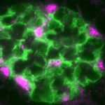
A master regulator of kidney health?
End-stage kidney disease often begins with injury to podocytes. These highly specialized cells are a critical part of the glomeruli, ...
-
Parsing the promise of inosine for neurogenic bladder
Spinal cord damage — whether from traumatic injury or conditions such as spina bifida — can have a profound impact on bladder ...
-
Modeling urinary tract disorders on a chip: Zohreh Izadifar
When a new tissue sample arrives from the Department of Urology, the Boston Children’s Hospital lab of Zohreh Izadifar, ...
-
A new tool could exponentially expand our understanding of bacteria
How do bacteria — harmless ones living in our bodies, or those that cause disease — organize their activities? A new study, ...