The Beauty of the Brain
Every year, the Harvard Brain Science Initiative sponsors its Beauty of the Brain contest. This year, two Boston Children’s Hospital images are among the six winners drawn from a pool of forty submissions.
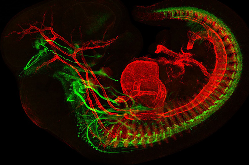
Above, Mary Whitman, MD, PhD, and Jess Bell, from the laboratory of Elizabeth Engle, MD, developed this image of a developing mouse embryo showing muscles in red and motor neurons in green. The Engle laboratory studies the genetics and molecular biology associated with eye disorders.
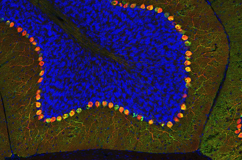
Below, Ellen DeGennaro, working in the laboratory of Christopher Walsh, MD, PhD, created this colorful image of a thin section of tissue from an adult mouse cerebellum. The image highlights blue nuclei, and Purkinje cells in red and green. The Walsh lab studies the genetics and molecular biology underlying human neurological diseases.
To view all the spectacular images visit the Beauty of the Brain
Related Posts :
-

Parsing the promise of inosine for neurogenic bladder
Spinal cord damage — whether from traumatic injury or conditions such as spina bifida — can have a profound impact on bladder ...
-
Unveiling the hidden impact of moyamoya disease: Brain injury without symptoms
Moyamoya disease — a rare, progressive condition that narrows the brain’s blood vessels — leads to an increased risk of stroke ...
-
Forecasting the future for childhood cancer survivors
Children are much more likely to survive cancer today than 50 years ago. Unfortunately, as adults, many of them develop cardiovascular ...
-
Genomic sequencing transforms a life: Asa’s story
Asa Cibelli feels like he’s been reborn. The straight-A middle schooler plays basketball and football, does jiu jitsu, is ...